Order Mermithida Hyman, 1951
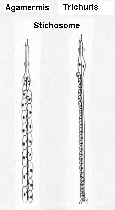
Diagnosis: Nematodes in this order are parasites of invertebrates. The main morphological characteristic of the order is the presence of a stichosome. Early juvenile stages with a protrusible stylet that is absent in the adults. Amphids dorylaimoid.
Family Mermithidae Braun, 1883
Diagnosis (Modified after Kaiser, 1991): Mermithoidea. Long, slender nematodes, usually between 10 and 100 mm. Cuticle smooth or containing criss-cross fibers near the outer layers, head containing two, four, or six cephalic papillae and sometimes a pair of lateral labial papillae. Oral opening terminal or shifted ventrally. Amphids tubelike or modified pouch-like. Esophagus, a stichosome usually with 4, 8, or 16 stichocytes. Intestine is modified as a food storage organ called the trophosome. Functional anus absent. Female with two ovaries; vagina muscular, straight or curved. Males with two testes. Spicules paired, fused, or single. Several rows of genital papillae usually present. Preparasitic juveniles with functional stylet.
Key to genera of terrestrial Mermithidae (Modified after Kaiser, 1991)
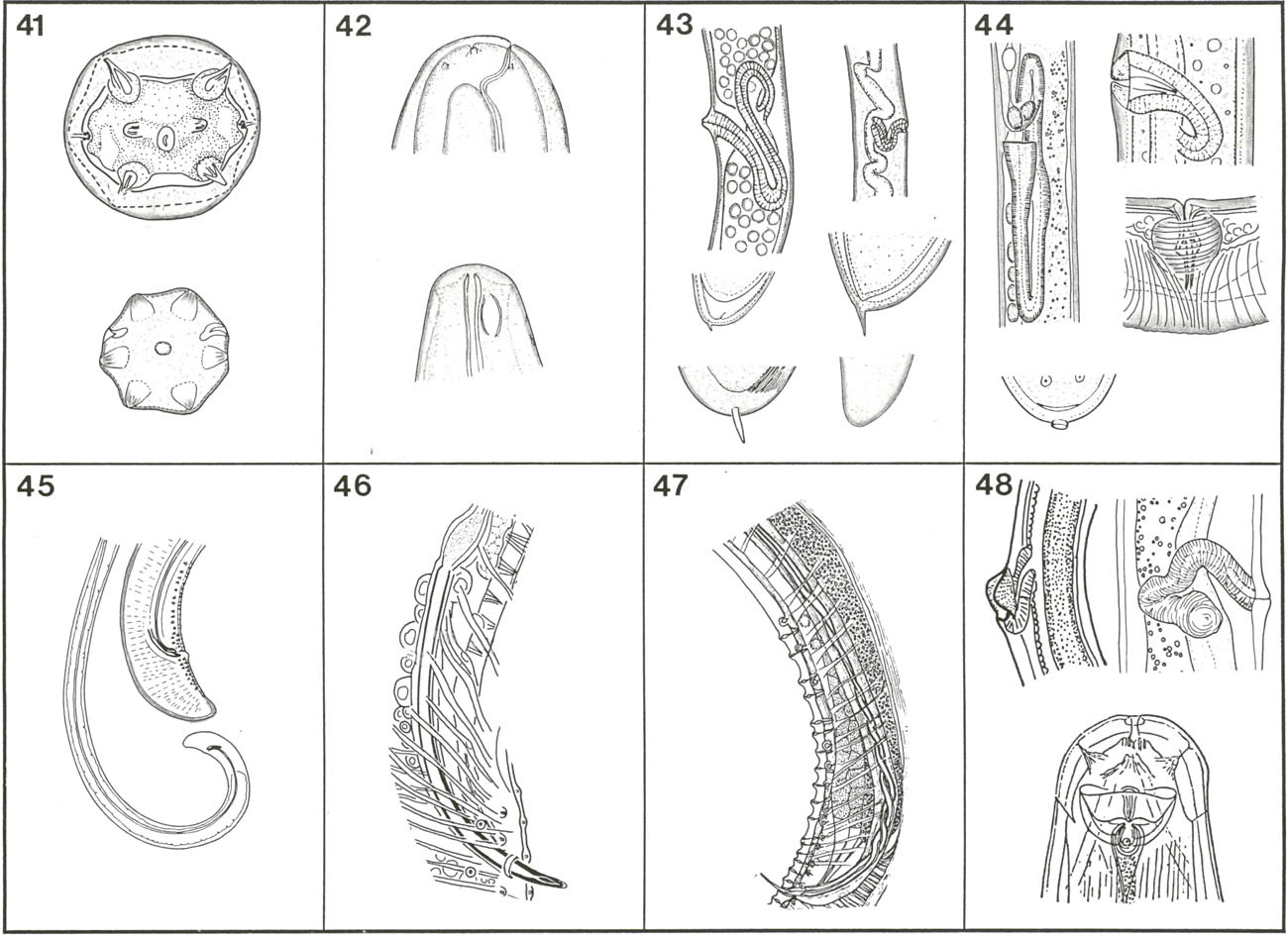
1- With four cephalic papillae (Fig. 41, top) 2
With six cephalic papillae (Fig. 41, bottom) 4
2- With two labial papillae (Fig. 41, top); oral opening terminal or slightly shifted to ventral side
(Fig. 42) Mermis Dujardin, 1842
Without labial papillae 3
3- Oral opening shifted ventrally as far posterior as the cephalic papillae; vagina
S-shaped (Fig. 43, left); postparasitic juveniles without tail appendage
Pheromermis Poinar, Lane and Thomas, 1976
Oral opening terminal, vagina short with V-like curve (Fig. 43, right); postparasitic
juveniles
with short tail appendage (Fig 43) Melolonthinimermis Artyukhovsky, 1963
4- Oral opening terminal; amphids small, cup-shaped with small pore-like aperture near the lateral cephalic papillae; vulva without flap, vagina with well-cuticularized lumen, horn- or S-shaped (Fig. 44, top right) 5
Oral opening, vagina, and amphids not as above 6
5- Parasitic and postparasitic juveniles with tail appendage
Hexamermis Steiner, 1924
Parasitic and postparasitic juveniles with a ring on tail end (Fig. 44, bottom); preparasitic juveniles lose the tail end during penetration of host
Agamermis Cobb, Steiner and Christie, 1923
6- Oral opening shifted slightly to ventral side; vagina modified S-shape (Fig. 44, top right) reflexed two or three times; spicules paired, one short and thick, the other extremely long and filiform (Fig. 45); postparasitic juvenile with short tail appendage Thaumamermis Poinar, 1981
Oral opening terminal or slightly shifted to ventral side; vagina and spicules not
as above 7
7- Oral opening shifted slightly ventrally, pharyngeal tube very long, can reach nine-tenths of body length; vulva oblique, vagina cylindrical, S-shaped; spicules long, parallel, with arch-like curve, 1.5-2 times of body diameter; parasitic and post parasitic juveniles with small tail appendage Oesophagomermis Artyukhovsky, 1969
Oral opening terminal 8
8- Vagina barrel-shaped (Fig. 44, right bottom) or cylindrical 9
Vagina S-shaped 10
9- Vagina short, barrel-shaped; spicules long, thin, parallel (Fig. 46), 2-5.5 times of body diameter; trophosome syncytial Psammomermis Polozhentsev, 1941
Vagina short but cylindrical and posteriorly directed; vulva flanked by lateral lips (Fig. 48, left); males unknown Tunicamermis Schuurmans-Stekhoven and Mawson, 1955
10- Spicules twisted (Fig. 47), parallel only in the midpart and on the distal end, fused on
tip Amphimermis Kaburaki and Imamura, 1932
Spicules parallel throughout their length 11
11- Amphids medium in size; vagina elongate, reflexed two or three times in two different planes (Fig. 48, right); spicules paired,
long, six or more times as long as body width
Aranimermis Poinar and Benton, 1986
Amphid very large (Fig. 48, bottom), vagina short but S-shaped; spicules short
Amphidomermis Filipjev, 1934
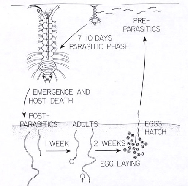
1- Aquatic mermithid: Romanomermis culicivorax
Infective juvenile (J2) swims in water seeking a suitable host. The nematode can survive in water for up to 48 hours. The body length is about 1mm. Upon contact , the nematode attaches to the host, using its stylet to make a hole on the cuticle and penetrates into the body cavity. The parasitic stage grows slowly for the first three or four days, and then rapidly increases in size. It takes 7 to eight days for the nematode to complete its cycle at 26C. In the later stages of development, the nematode can be seen in side the host, either coiled around the thorax or fold longitudinally along the body. The nematode then molts to post parasitic stage, ruptures the host cuticle and emerges. This emergence is always fatal to the larvae host. For some other mermithids the nematode develops further until the host becomes adult.
After emergence, the nematodes (some time called postparasitic nematodes) burrow in the soil where they become adult in 7 or more days. The adults mate and the females begin to lay eggs. The number of eggs is variable depending on species of the nematodes. For Romanomermis culicivorax, the female lays about 2500 eggs over a period of 10-15 days. Usually, the life cycle is completed in 3-6 weeks.
Life cycle of mermithid
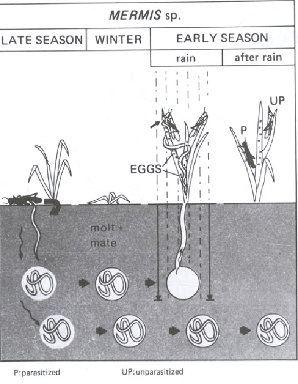
2- Terrestrial mermithid: Mermis nigrescens
The fully developed parasitic juveniles leave their host and migrate into the soil. They penetrate to the depth of 20-40 cm, depending upon the degree of moisture needed to survive. The juveniles molt into adults. After copulation, the females begin to produce eggs. The eggs are retained in the uterus, where the embryonic development takes place and where they are provided with an additional protection against dehydration. The eggs of the species have polar hairs to hold the eggs onto the vegetation after being laid. After rainfall in the early season, the adult females that contact water, move out of soil, creep slowly over the vegetation laying their eggs. Females that are not affected by water remain in the soil until heavier rains cause them to emerge. When the insects, as grasshoppers, feed on vegetation, they ingest mermithid eggs. Infective juveniles hatch in insect intestine, then penetrate the intestinal wall into body cavity, where they grow to a length of 9 to 15 cm in 4 to 10 weeks.
3- Terrestrial mermithid: Amphimermis, Agamermis, Hexamermis.
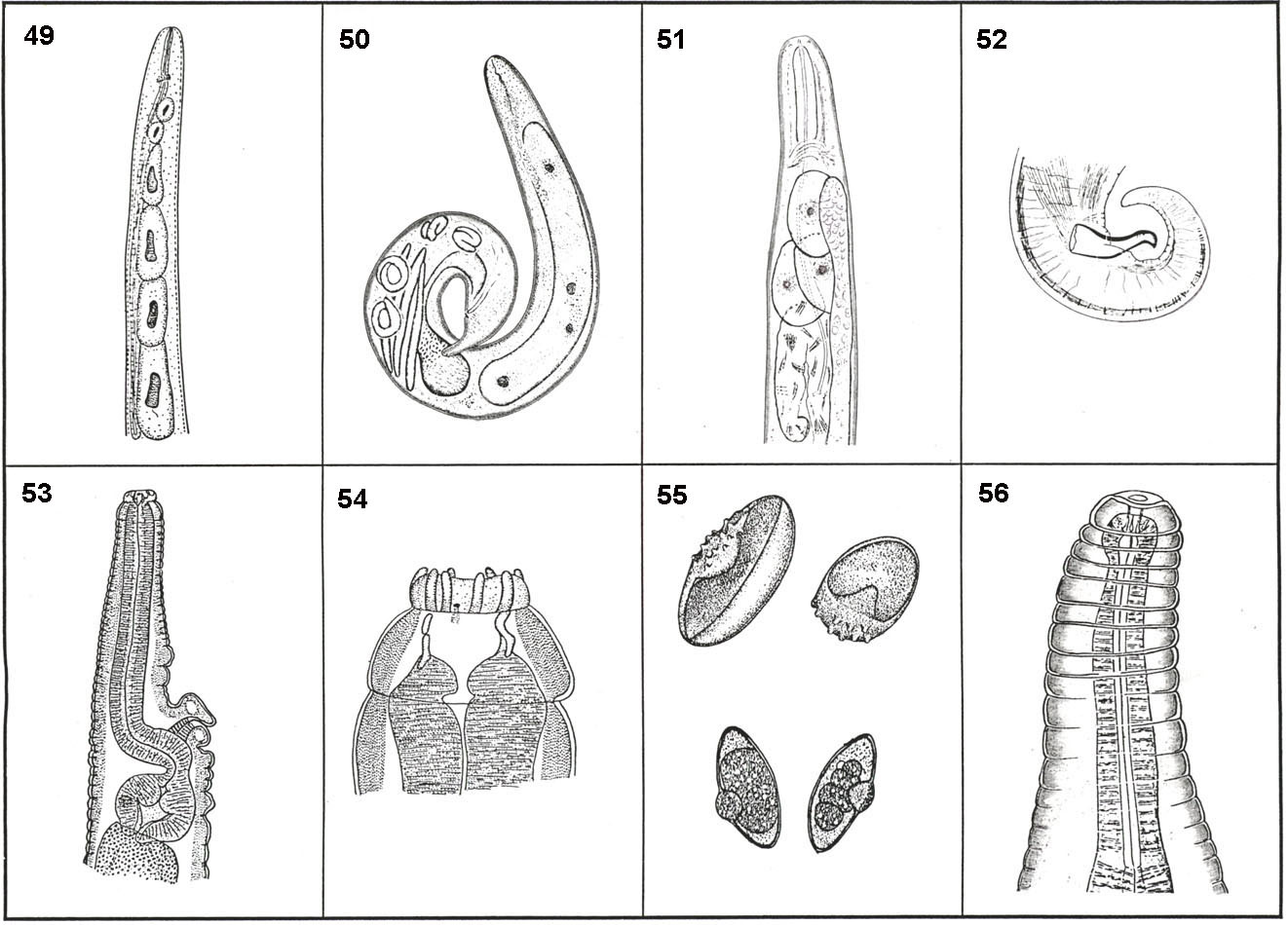
The cycle of these nematodes are closer to that of Mermis but instead of the females move out of soil and lay eggs on vegetation, the eggs hatch in soil and the infective juvenile move out and seek for a new host. The juveniles (J2) bore through the body wall into the body cavity.
Family Tetradonematidae Cobb, 1919
Diagnosis: (After Poinar, 1975). Mermithoidea. Lips rudimentary or absent; juveniles with a minute stylet which disappears in later stages; head papillae reduced; esophagus consists of a simple hollow tube and associated tetrad (four-celled structure as in Fig. 49); anus absent; ovaries and testes paired; vulva mid-body; spicule single, bursa absent. The nematodes mature to the adult stage and mate in the body cavity of the insect host.
Key to the genera of
Tetradonematidae (After Poinar, 1975)
1- Life cycle with two parasitic generations; first generation parthenogenetic with
female lacking a vulva; males of second generation producing both sperm and eggs (Fig. 50)
Heterogonema van Waerebeke and Remillet, 1971
Life cycle with single parasitic generation; female with vulva; male producing
sperm only 2
2- Pharyngeal region of the female containing a tetrad (four large cells arranged in a row as in Fig. 49); mature female with an acute tail; male tail lacking genital papillae …………………………………………….. .Tetradonema Cobb, 1919
Pharyngeal region of the female not containing a tetrad; mature female with a
round tail; male with or without genital papillae 3
3- Male lacking genital papillae Aproctonema Keilin, 1917
Male possessing genital papillae 4
4- Mature forms with three large gland-like cells posterior to the nerve-ring (Fig. 51), stichosome and stichocytes lacking; spicule of male with tip curved downward (Fig. 52) Corethrellonema Nickle, 1969
Mature forms lacking three large cells behind the nerve ring; stichosome with eight
stichocytes present in the adults; spicule of male not curved downward at tip
Mermithonema Goodey, 1941
Update on June 2011