Order Oxyurida
Weinland, 1858
(in Yamaguti, 1961) (syn.: Oxyurata Skrjabin, 1923, in Skryabin et al., 1951)
Diagnosis: Small to medium nematodes, obligatory zooparasites. Eight papillae present in one circle, sometimes they fuse to form four papillae. Amphids small. Stoma cylindrical but short, not cuticularized, not surrounded by esophageal tissue posteriorly. Esophagus with corpus, isthmus and basal bulb; basal bulb valvate. Corpus is either cylindrical, fusiform or composed of procorpus and metacorpus. Female gonads monodelphic or didelphic, terminal portion muscular. Eggs usually with relatively thin shell, and mostly do not hatch until ingested by an appropriate host. Male with one, two, or no spicules. Posterior portion of male with genital papillae; bursa usually present.
This order has two superfamilies: Thelastomatoidea, parasites of invertebrates, and Oxyuroidea, parasites of vertebrates. The superfamily Thelastomatoidea was studied taxonomically by Adamson (1989), and Adamson and Waerebeke (1992a, 1992b, 1992c). They divided the nematodes in this order into five families: Thelastomatidae, Travassosinematidae, Protrelloididae, Pseudonymidae, and Hystrignathidae, and gave the descriptions of genera for each family. Because of space limitations, and lack of information, we will mention only three families, Thelastomatidae, Hystrignathidae, and Travassosinematidae.
Family Thelastomatidae Travassos, 1929
Diagnosis: Thelastomatoidea. Anterior end with eight papillae. Amphid rounded or oval. Esophagus with variable corpus, distinct or indistinct isthmus, and a valvular basal bulb. Females with one or two gonads; vulva anterior or posterior to base of esophagus. Male with single spicule or none; tail with 1-4 genital papillae.
Key to common genera, parasites of Blattaria (Cockroaches)
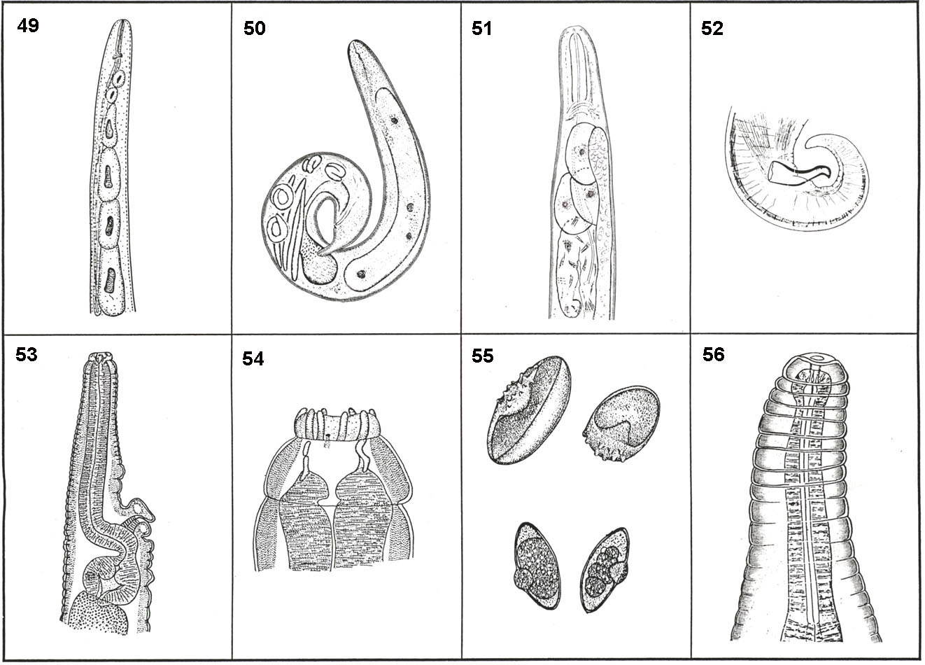
1- Vulva anterior to posterior end of esophagus (Fig. 53) 2
Vulva posterior to posterior end of esophagus 4
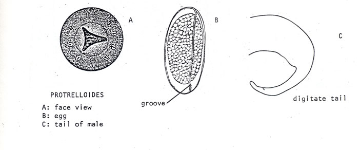
2- Esophagus of female long, slender, about 1/4 of
body length; male tail digitate Protrelloides Chitwood, 1932
Esophagus not long and slender; male tail not digitate 3
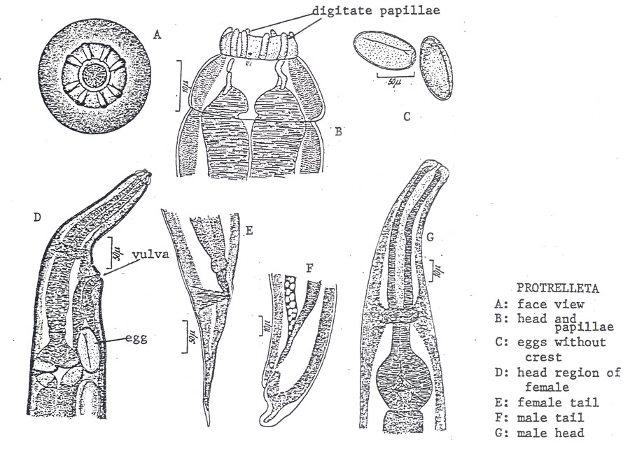
3- Lip region bearing eight prominent digitate papillae (Fig. 54), eggs without cuticular crest; no
spicules in male Protrelleta Chitwood, 1932
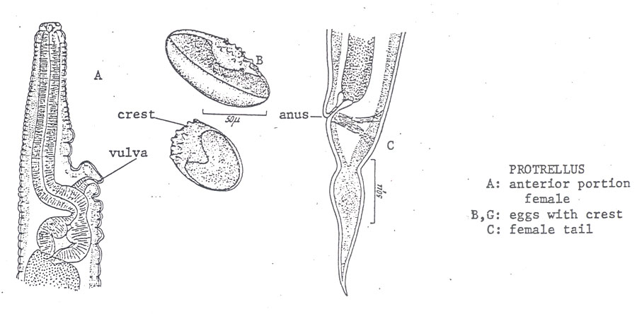
Lip region not bearing eight prominent digitate papillae, eggs with cuticular
crest (Fig. 55), one spicule in male Protrellus Cobb, 1920
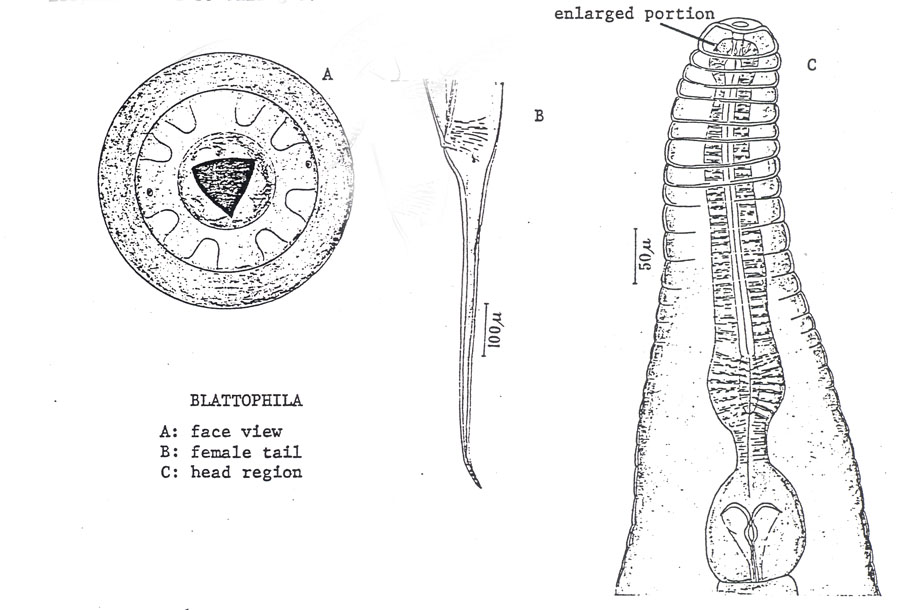
4- Esophagus enlarged subspherically at base of buccal cavity (Fig. 56)
Blattophila Cobb, 1920
Esophagus not so enlarged 5
5- Esophagus with median bulb 6
Esophagus without median bulb 9
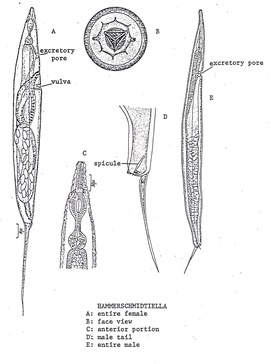
6- Vulva in anterior part of the body, 1/4-1/3 body length from
anterior end, Hammerschmidtiella Chitwood, 1932
Vulva in mid or posterior part of the body, 2 or more body lengths
from anterior end 7
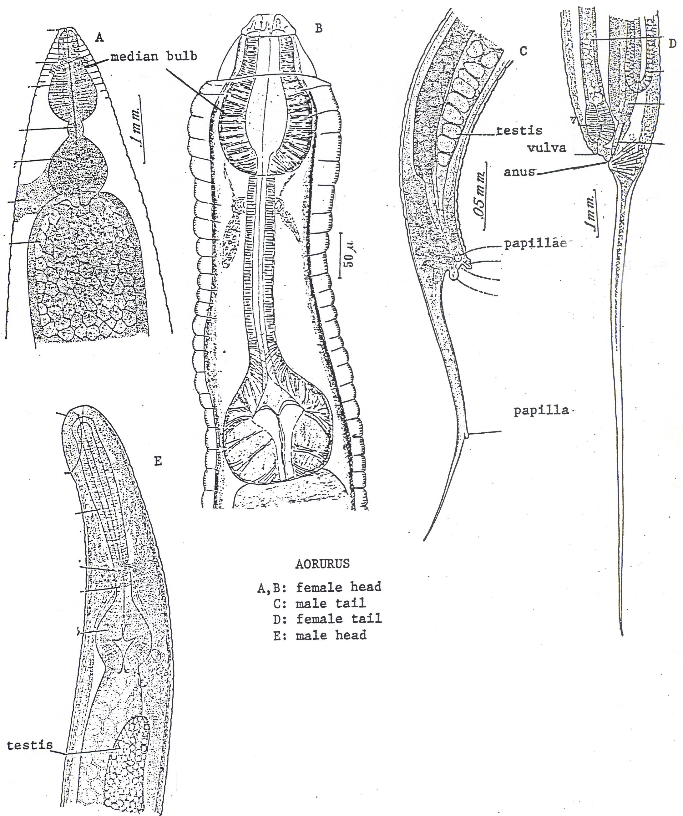
7- Anterior part of esophagus very short, median bulb large (Fig. 57),
pear-shaped Aorurus Leidy, 1849
Anterior part of esophagus long, median bulb not as above 8
8- Median bulb cylindrical (Fig. 58) Leidynema Schwenk in Travassos, 1929
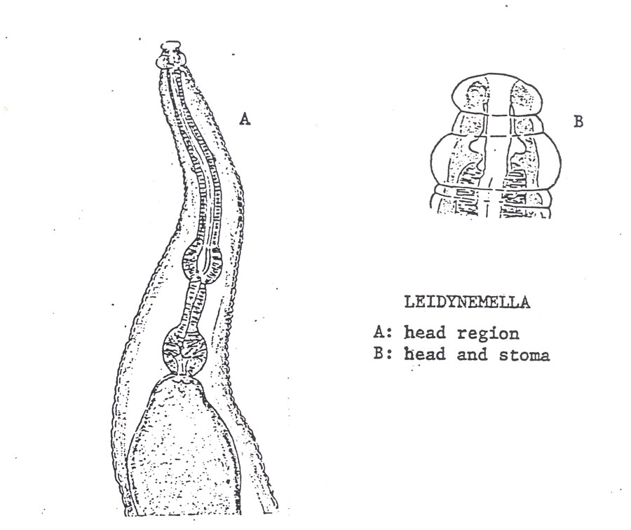
Median bulb spherical (Fig. 59) Leidynemella Chitwood and Chitwood, 1934
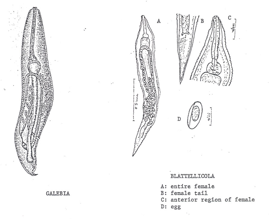
9- Female with one gonad 10
Female with two gonads 12
10- Esophagus long, about 1/3 body length, tail short (Fig. 60)
Galebia Chitwood, 1932
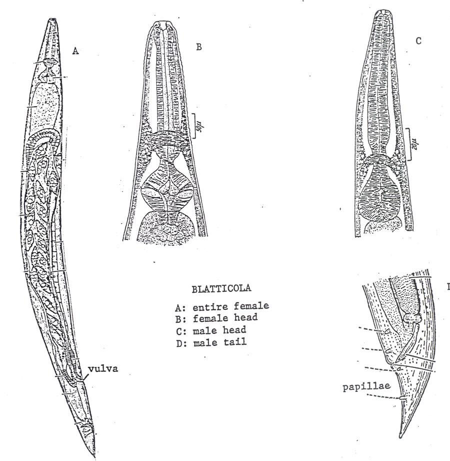
Esophagus not long, about 1/6 or less body length 11
11- Female tail attenuate, vulva located in middle 1/3 of body
Blatellicola Basir, 1940
Female tail conical, vulva located in posterior 1/3 of body
Blatticola Schwenk, 1926
12- Eggs fused in pairs along flattened surfaces (Fig. 61) Cameronia Basir, 1948
Eggs not fused 13
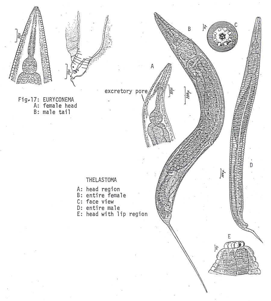
13- Female tail filiform 14
Female tail not filiform (Fig. 62, left) 15
14- Excretory pore of female present, male tail filiform or
delicately attenuated (Fig. 62, right) Thelastoma Leidy, 1849
Excretory pore of female not observed, male tail very short,
degenerate Euryconema Chitwood, 1932
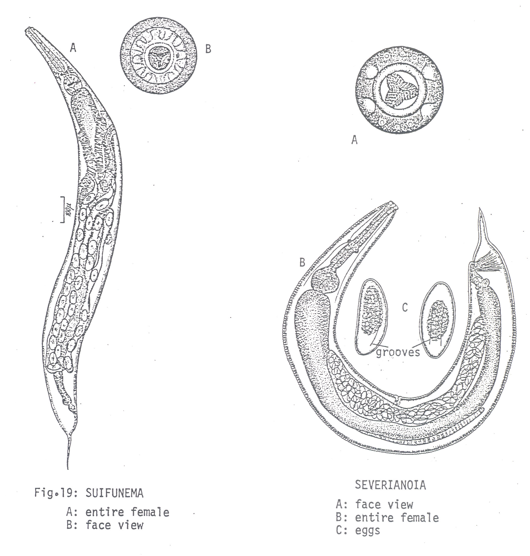
15- Vulva anterior to mid-body Suifunema Chitwood, 1932
Vulva near mid-body 16
16- Eggs with longitudinal grooves, excretory pore not observed (Fig. 63)
Severianoia (Schwenk, 1926) Travassos, 1929
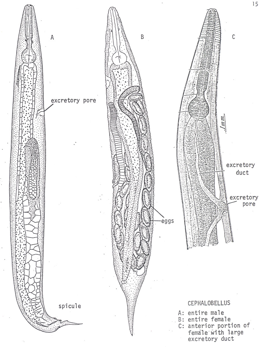
Eggs without longitudinal grooves, excretory pore posterior to basal bulb, excretory duct
very large (Fig. 64) Cephalobellus (Syn. Scarabanema) Cobb, 1920
Notes: 1- Cameronia is a parasite of mole crickets. 2- Even though we have included Blattellicola in the key, this genus was synonimized with Blatticola by Adamson and Waerebeke (1992a).
Family Hystrignathidae Travassos, 1920
Diagnosis: Thelastomatoidea. Cuticle with or without spines or
scales. Anterior end with eight papillae. Amphids small. Cephalic region with
one or two enlarged annules. Esophageal corpus either cylindrical or divided
with posterior part clavate. Isthmus well defined or indistinct. Basal bulb
mostly with valves. Females with one or two gonads. Eggs elongate, ornamented
with ridges or excrescences. Tail conical, attenuate, sometimes with rounded
terminus or with appendage. Male mostly absent, when observed, spicule either
single or absent; at least one median single papilla observed. Nematodes in
this family are parasites of beetles in family Passalidae.
Key to genera of Hystrignathidae
1- Anterior region with transverse, or longitudinal rows of spines 2
Anterior region without rows of spines 12
2- Female with one gonad 3
Female with two gonads 6
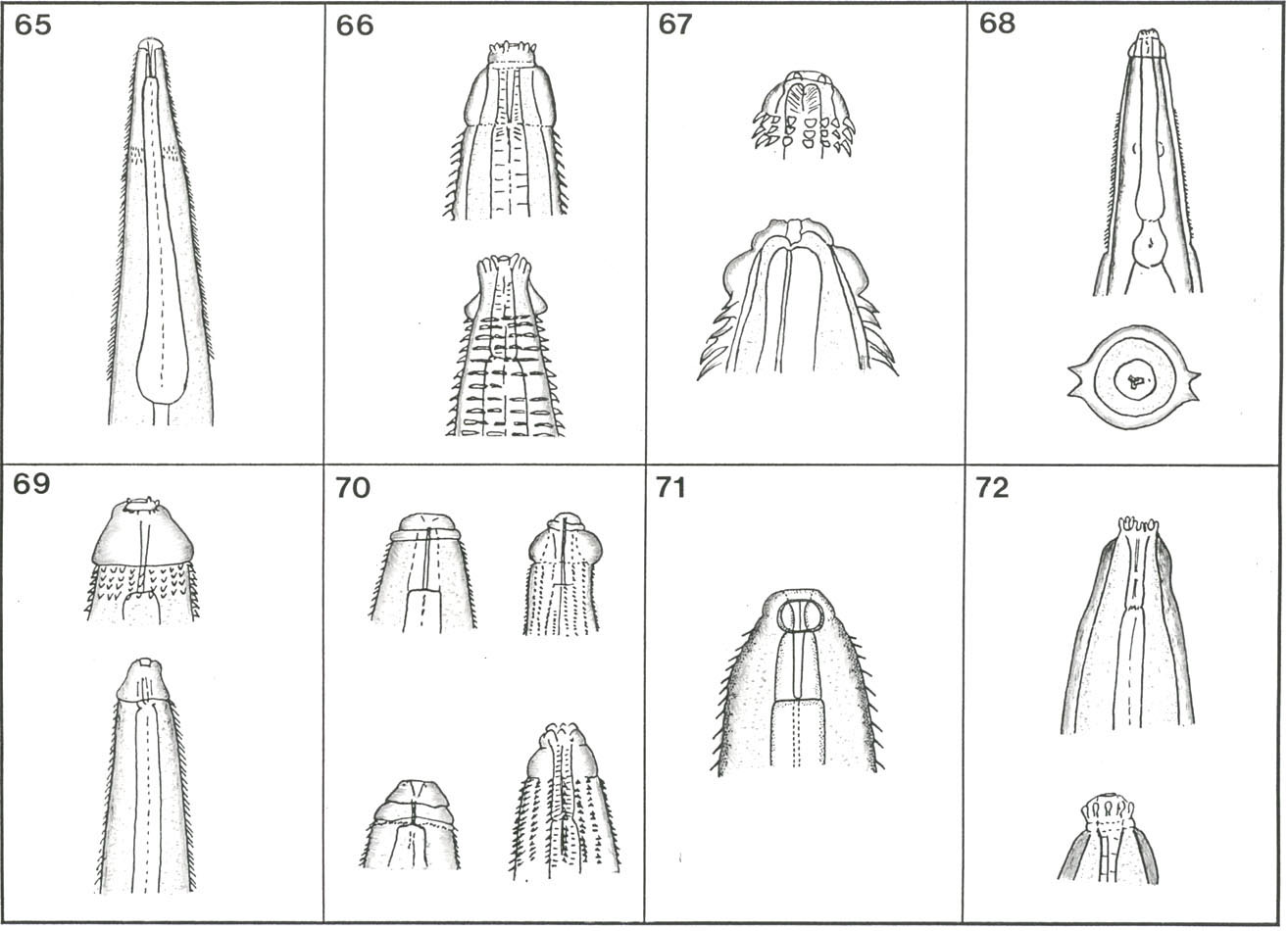
3- Corpus cylindrical anteriorly, clavate posteriorly (Fig. 65) … Artigasia Christie, 1934
Corpus cylindrical without clavate portion 4
4- Spines in successive rows alternating Boraceianema Travassos and Kloss, 1958
Spines in successive rows, aligned 5
5- Oral opening surrounded by pedunculate papillae (Fig. 66)
Klossiella Cordeiro, 1981
Oral opening not surrounded by pedunculate papillae
Mentecle Travassos and Kloss, 1958
6- Corpus cylindrical without clavate posterior portion 7
Corpus cylindrical anteriorly, clavate posteriorly 8
7- Cervical cuticle with transverse rows of scale-like projections (Fig. 67); male
present Lepidonema Cobb, 1898
Cervical cuticle with transverse rows of spines, male unknown
Soaresnema Travassos and Kloss, 1958
8- Cervical cuticle with two lateral longitudinal rows of spines (Fig. 68)
Carlosia Travassos and Kloss, 1957
Cervical cuticle with transverse rows of spines 9
9- Cervical cuticle with transverse rows of scale-like projections, cephalic region
with single circumoral annule (Fig. 69) Salesia Travassos and Kloss, 1958
Cervical cuticle with transverse rows of spines, cephalic region with two annules
(Fig. 70) 10
10- First row of spines with 16 elements Hystrignathus Leidy, 1850
First row of spines with 32 elements 11
11- Buccal cavity divided into anterior spheroid and posterior cylindrical segments
(Fig. 71) Urbanonema Travassos and Kloss, 1958
Buccal cavity without anterior spheroid chamber Xyo Cobb, 1898
12- Female with one gonad 13
Female with two gonads 17
13- Anterior end
with eight pedunculate papillae (Fig. 72)
Coronocephalus Cordeiro, 1981
Anterior end without pedunculate papillae 14
14- Two cephalic annules contiguous Glaber Travassos and Kloss, 1958
Two cephalic annules separate 15
15- Body robust, fusiform Passalidophila Van Waerebeke, 1973
Body neither robust nor fusiform 16
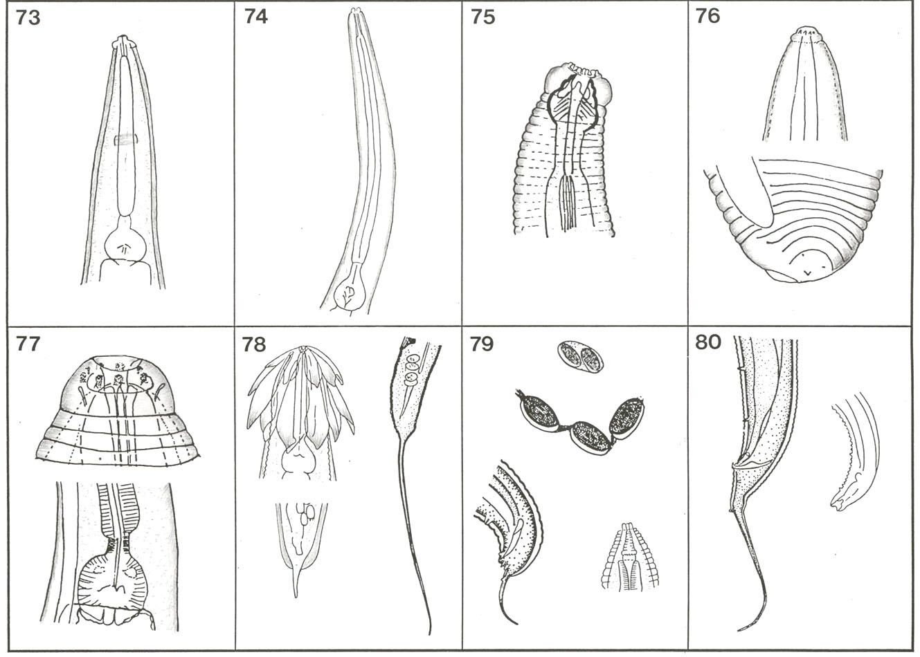
16- Esophageal corpus short, spindle-shaped (Fig. 73)
Christiella Travassos and Kloss, 1957
Esophageal corpus long, cylindrical (Fig. 74) Longior Travassos and Kloss, 1958
17- Esophageal corpus with posterior portion not clavate 18
Esophageal corpus with posterior portion clavate 19
18- Anterior end of esophagus swollen, surrounding base of stoma (Fig. 75)
Anomalostoma Cordeiro, 1981
Anterior end of esophagus not as above Ventelia Travassos and Kloss, 1958
19- Cephalic end with two annules, tail short, rounded with mucron-like appendage
(Fig. 76) Anuronema Clark, 1978
Cephalic end with one annule, tail not as above 20
20- Circumoral annule in form of truncate cone, isthmus well-defined (Fig. 77)
Phalacronema Clark, 1978
Circumoral annule not as above, isthmus mostly ill-defined 21
21- Vulva in posterior quarter of body Klossnema Cordeiro, 1981
Vulva near mid-body 22
22- Lateral alae broad, bulb without valves Triumphalisnema Kloss, 1962
Lateral alae not broad, bulb with poorly developed valves Sprentia Clark, 1978
Family Travassosinematidae Rao, 1958
Diagnosis: Head with or without leaflike extension. Eggs included in a membrane either connected with each other or separate with one, two or three eggs in a capsule. Male without spicules except in Binema and Isobinema. Most nematodes in this family are parasites of mole crickets.
Key to genera of Travassosinematidae
1- Female head with leaf-like extensions (Fig. 78, top left) 2
Female head without leaf-like extensions 4
2- Body with rows of spines
Body without rows of spines 3
3- Females with short tail (Fig. 78, bottom) Pulchrocephala Travassos, 1925
Females with long, filiform tail (Fig. 78, right) Travassosinema Rao, 1958
4- Eggs in capsules of two or three (Fig. 79) 5
Eggs attached together in a chain (Fig. 79) 6
5- Male with small spicule (Fig. 79, bottom left) Binema Travassos, 1925
Male without spicule Mirzaiella Basir, 1942
6- Stoma long, annulated (Fig. 79, bottom right) Chitwoodiella Basir, 1948
Stoma not long, not annulated 7
7- Male tail flagellate (Fig. 80, left) Isobinema Rao, 1958
Male tail truncate (Fig. 80, right) Singhiella Rao, 1958
____________________
Update April 19 2010